1 / 5
| Model | Sheath O.D.D±0.1(mm) | Working Length L±50(mm) | Needle Size (Diameter/Length) | Endoscopic Channel (mm) |
| ZRH-PN-2418-214 | Φ2.4 | 1800 | 21G,4mm | ≥2.8 |
| ZRH-PN-2418-234 | Φ2.4 | 1800 | 23G,4mm | ≥2.8 |
| ZRH-PN-2418-254 | Φ2.4 | 1800 | 25G,4mm | ≥2.8 |
| ZRH-PN-2418-216 | Φ2.4 | 1800 | 21G,6mm | ≥2.8 |
| ZRH-PN-2418-236 | Φ2.4 | 1800 | 23G,6mm | ≥2.8 |
| ZRH-PN-2418-256 | Φ2.4 | 1800 | 25G,6mm | ≥2.8 |
| ZRH-PN-2423-214 | Φ2.4 | 2300 | 21G,4mm | ≥2.8 |
| ZRH-PN-2423-234 | Φ2.4 | 2300 | 23G,4mm | ≥2.8 |
| ZRH-PN-2423-254 | Φ2.4 | 2300 | 25G,4mm | ≥2.8 |
| ZRH-PN-2423-216 | Φ2.4 | 2300 | 21G,6mm | ≥2.8 |
| ZRH-PN-2423-236 | Φ2.4 | 2300 | 23G,6mm | ≥2.8 |
| ZRH-PN-2423-256 | Φ2.4 | 2300 | 25G,6mm | ≥2.8 |
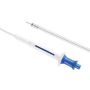
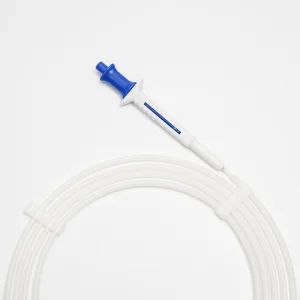
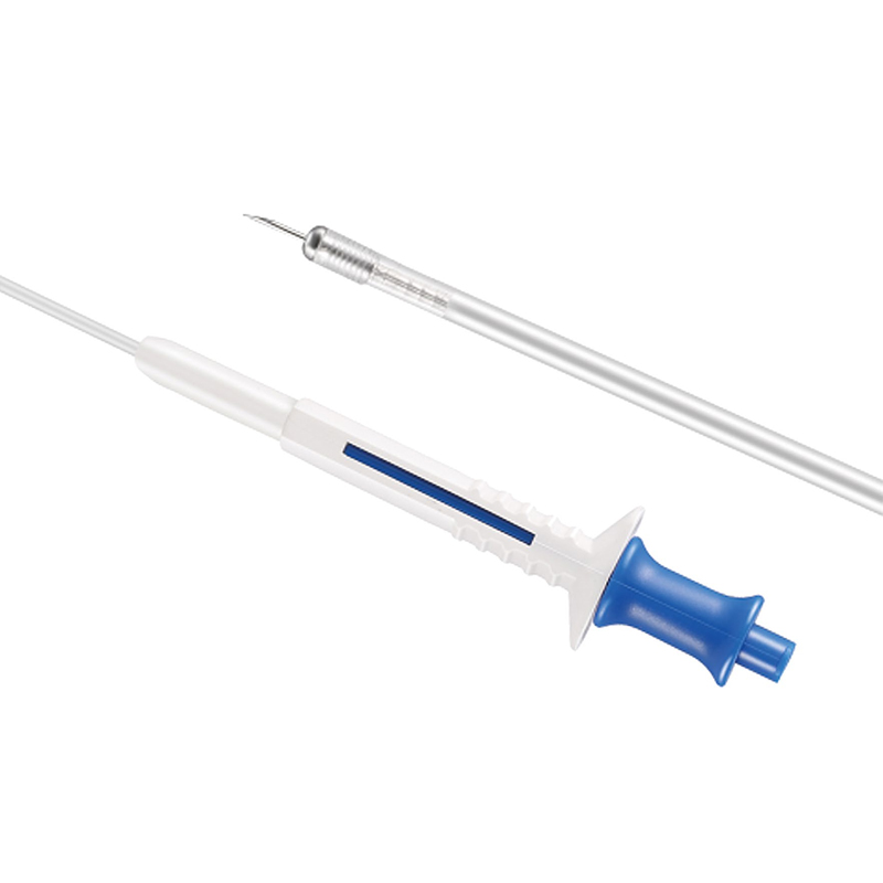
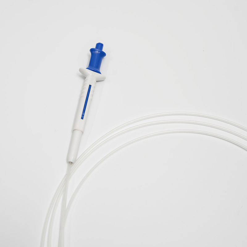
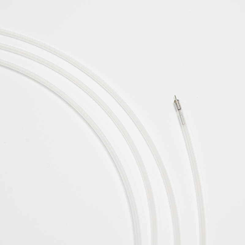
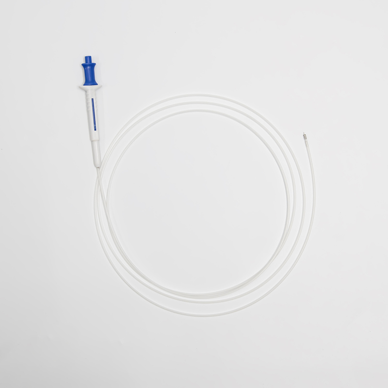
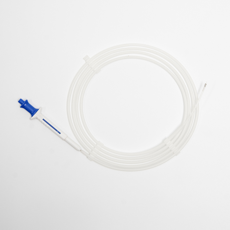
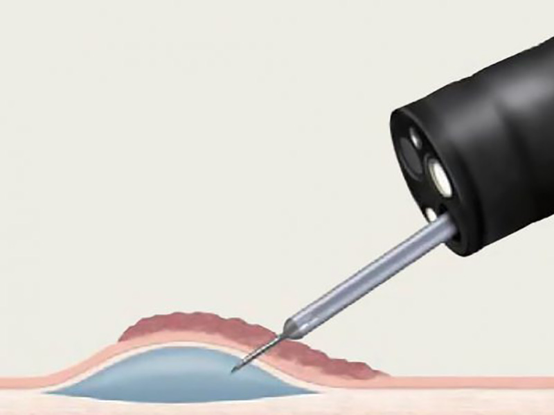
A sclerotherapy needle is used to inject fluid into the submucosal space to elevate the lesion away from the underlying muscularis propria and create a less flat target for resection.
Can be used to observe blood return.
Facilitates advancement through difficult pathways.
Sharp puncture
Easy to control the needle moving.
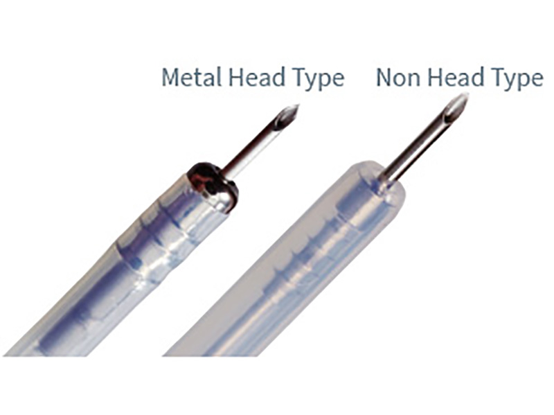
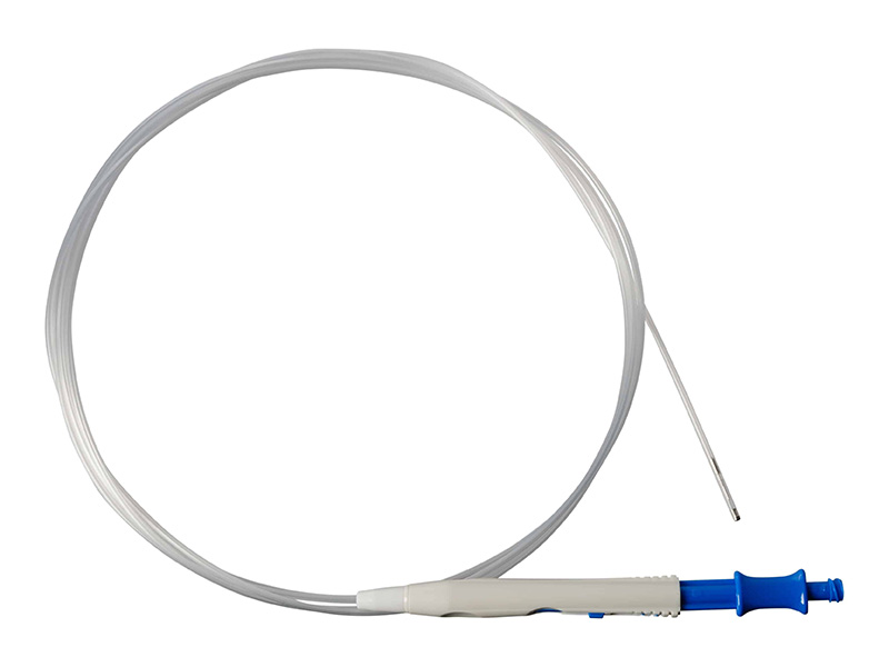
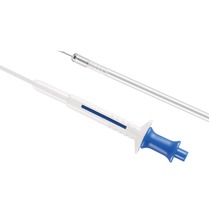
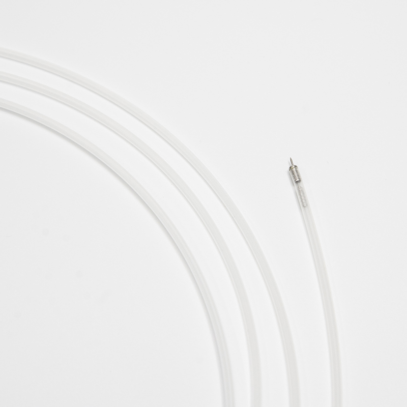
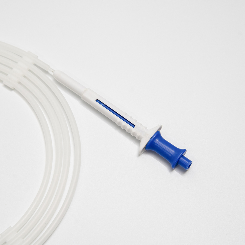
A sclerotherapy needle is used to inject fluid into the submucosal space to elevate the lesion away from the underlying muscularis propria and create a less flat target for resection. The injection is often done with saline, but other solutions have been used to achieve longer maintenance of the bleb including hypertonic saline (3.75% NaCl), 20% dextrose, or sodium hyaluronate [2]. Indigo carmine (0.004%) or methylene blue is often added to the injectate to stain the submucosa and provides a better evaluation of the depth of resection.
The submucosal injection can also be used to determine if a lesion is appropriate for endoscopic resection. Lack of elevation during injection indicates adherence to the muscularis propria and is a relative contraindication to proceeding with EMR. After creating the submucosal elevation, the lesion is grasped with a rat tooth forceps that has been passed through an open polypectomy snare. The forceps lifts the lesion and the snare is pushed down around its base and resection ensues. This “reach-through” technique requires a double lumen endoscope which can be cumbersome to use in the esophagus. As a result, lift-and-cut techniques are used less commonly for esophageal lesions.
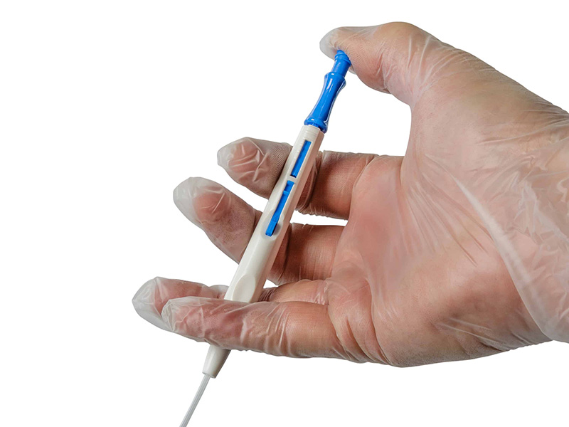
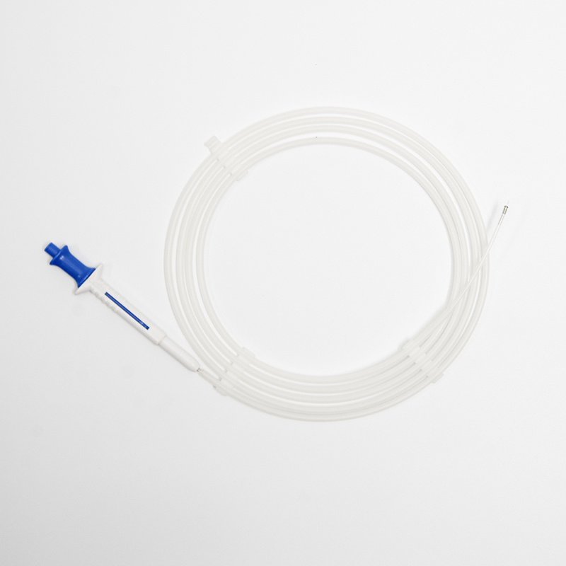
It is primarily intended for endoscopic injection of sclerotherapy agents and dyes into esophageal or colonic varices, injecting saline to aid in EMR and polypectomy procedures, and controlling non-variceal haemorrhage.
The needles are available in 21G, 23G, and 25G diameters with needle projection lengths of either 4mm or 6mm, compatible with working lengths of 1800mm and 2300mm.
All listed models have a sheath outer diameter of Φ2.4 mm and require an endoscopic channel size of ≥2.8 mm.
The transparent inner tube allows the operator to clearly observe blood return, ensuring precise placement and safety during the injection procedure.
Injecting fluid elevates the lesion away from the underlying muscularis propria, creating a protruding target that facilitates safer and more effective snare resection while reducing perforation risk.
A lack of elevation often indicates tissue adherence or invasion into the muscularis propria, serving as a relative contraindication to proceeding with standard EMR.